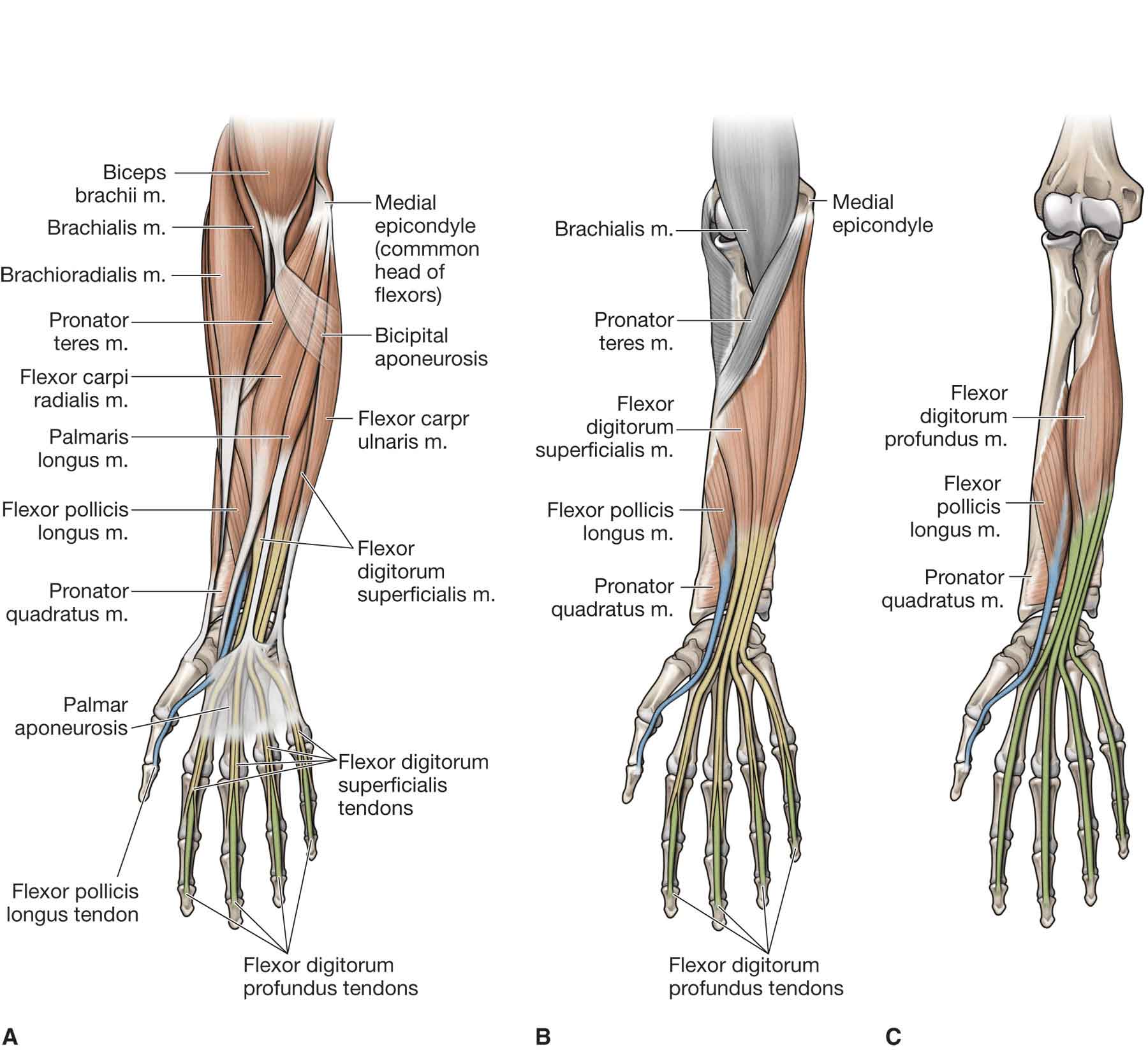
Picture Of Forearm Muscles And Tendons / Posterior Forearm | Basicmedical Key. A deep layer , intermediate layer and superficial layer. Long flexor tendons extend from the forearm muscles through the wrist and attach to the small bones of the fingers and thumb. These types of strains are quite severe and involve complete rupture of the muscle fibers and tendons. By moving the mouse cursor over a particular area of the arm or forearm, this area is highlighted and the labels are displayed: While the ventral side of the forearm is not exactly less complicated than the dorsal side, it appears less complicated on the.
A square shaped muscle found deep to the tendons of the fdp and fpl. The superficial group arises mostly from the posterior aspect of the lateral epicondyle of the humerus by a common tendon. Lesson on the anatomy of the forearm: These types of strains are quite severe and involve complete rupture of the muscle fibers and tendons. Two of the tendons combine visually so the end result looks like two thick tendons, with a long hollow between them.
Wrist Tendons Anatomy - Anatomy Diagram Book from musculoskeletalkey.com When identifying the function of the forearm muscles, it is important to note that any forearm compartment muscle that crosses the elbow joint will act at this joint. From superior to inferior, origin. Stretching is a good way to strengthen your forearm muscles and release any pain or stress. Resting the muscles in the affected tendons is crucial to treating tendinitis, especially in athletes. It is separated from the anterior compartment by the interosseous membrane between the radius and ulna. Grade i strain of forearm muscle: Two of the tendons combine visually so the end result looks like two thick tendons, with a long hollow between them. Important questions on muscles of flexor and extensor compartment of forearm, structures deep and superficial to 3 name the muscles of flexor compartment of forearm supplied by median nerve.
Find stockbilleder af forearm muscles tendons i hd og millionvis af andre royaltyfri stockbilleder, illustrationer og vektorer i shutterstocks samling.
In the anterior compartment, they are split into three categories: Tusindvis af nye billeder af høj kvalitet tilføjes hver dag. Lesson on the anatomy of the forearm: It is separated from the anterior compartment by the interosseous membrane between the radius and ulna. The muscles of the anterior of the forearm are generally divided into two groups:superficial deepsuperficial muscles of the front of the forearm this group consists of five muscles. The muscle passes obliquely across the forearm, and ends in a flat tendon, which is inserted into a rough impression at the middle of the lateral surface the flexor carpi radialis lies on the medial side of the preceding muscle. From superior to inferior, origin. Also, pollicis means thumb in latin. There are many muscles in the forearm. Stretching is a good way to strengthen your forearm muscles and release any pain or stress. Grade i strain of forearm muscle: The ecrb tendon is one of 3 tendons, including ecrl and ecu, which act together to bend back the wrist. Hold your elbow with thumbs up and other 4 extension of index finger.
These types of strains are quite severe and involve complete rupture of the muscle fibers and tendons. Do it yourself as shown in the picture! A constant stretching and strengthening routine can help to alleviate forearm tendinitis. The supinator is a muscle. If you keep your hand flat on a table and.
human forearm tendon detail - Google Search | SDP Tendons and Forces | Pinterest | Anatomy ... from s-media-cache-ak0.pinimg.com Find stockbilleder af forearm muscles tendons i hd og millionvis af andre royaltyfri stockbilleder, illustrationer og vektorer i shutterstocks samling. Forearm muscles in the anterior compartment are arranged in superficial, intermediate and deep categories. Resting the muscles in the affected tendons is crucial to treating tendinitis, especially in athletes. It turns… inflamed common flexor tendon cft. Grade i strain of forearm muscle: A deep layer , intermediate layer and superficial layer. The posterior compartment of the forearm (or extensor compartment) contains twelve muscles which are chiefly responsible for extension of the wrist and digits, and supination of the forearm. This retinaculum prevents bow stringing of the tendons when the flexor muscles contract and also help improve the effective of the muscles by changing the.
Its muscle belly is in the forearm and then travels to the thumb side of the wrist on the back part of the forearm.
While the ventral side of the forearm is not exactly less complicated than the dorsal side, it appears less complicated on the. From superior to inferior, origin. Anconeus muscle is a small muscle that is triangular in shape. The muscle passes obliquely across the forearm, and ends in a flat tendon, which is inserted into a rough impression at the middle of the lateral surface the flexor carpi radialis lies on the medial side of the preceding muscle. Originates from the anterior surface of the ulna and attaches to the. The ecrb tendon is one of 3 tendons, including ecrl and ecu, which act together to bend back the wrist. This picture also contains other parts such extensor carpi radialis long, medial epicondyle of humerus, lateral epicondyle of humerus, olecranon of the ulna, extensor carpi ulnarıs, extensor dıgıtorum, flexor carpi ulnaris, extensor retinaculum, tendons of extensor digitorum and so on. When identifying the function of the forearm muscles, it is important to note that any forearm compartment muscle that crosses the elbow joint will act at this joint. Long flexor tendons extend from the forearm muscles through the wrist and attach to the small bones of the fingers and thumb. Forearm pain from muscle or tendon injuries can be quite debilitating. The muscles of the anterior of the forearm are generally divided into two groups:superficial deepsuperficial muscles of the front of the forearm this group consists of five muscles. Important questions on muscles of flexor and extensor compartment of forearm, structures deep and superficial to 3 name the muscles of flexor compartment of forearm supplied by median nerve. All 4 muscles have a common origin at the medial epicondyle of the humerus, known as the common flexor tendon.
Tendons are attached to muscles and to bone. The ecrb tendon is one of 3 tendons, including ecrl and ecu, which act together to bend back the wrist. See anatomy pictures of the 27 bones in the hand and wrist, how they are connected with tendons and muscles and the nerves that run through the skeletal structure. These types of strains are quite severe and involve complete rupture of the muscle fibers and tendons. Stretching is a good way to strengthen your forearm muscles and release any pain or stress.
6: Upper Limb | Basicmedical Key from basicmedicalkey.com Learning their anatomy will help you design awesomely dynamic arms. This type of forearm grade iii strain of forearm muscle: The muscle passes obliquely across the forearm, and ends in a flat tendon, which is inserted into a rough impression at the middle of the lateral surface the flexor carpi radialis lies on the medial side of the preceding muscle. It turns… inflamed common flexor tendon cft. Originates from the anterior surface of the ulna and attaches to the. The anterior forearm muscles are divided into 3 muscular layers ; In the anterior compartment, they are split into three categories: Find stockbilleder af forearm muscles tendons i hd og millionvis af andre royaltyfri stockbilleder, illustrationer og vektorer i shutterstocks samling.
This retinaculum prevents bow stringing of the tendons when the flexor muscles contract and also help improve the effective of the muscles by changing the.
Muscles of the forearm segregate into these compartments consisting of (1) an anterior group (the flexors seven superficial and five deep muscles occupy the posterior forearm. The extensor digitorum is a muscle belly, passing first into four tendons, which in turn transformirovalsya in stretching the tendon fixed to the base of the distal. All superficial muscles are arises from the medial epicondyle of humerus but they are inserted into the different part except. Two of the tendons combine visually so the end result looks like two thick tendons, with a long hollow between them. Stretching is a good way to strengthen your forearm muscles and release any pain or stress. While the ventral side of the forearm is not exactly less complicated than the dorsal side, it appears less complicated on the. It's sometimes blended with triceps brachii or extensor carpi ulnaris. This picture also contains other parts such extensor carpi radialis long, medial epicondyle of humerus, lateral epicondyle of humerus, olecranon of the ulna, extensor carpi ulnarıs, extensor dıgıtorum, flexor carpi ulnaris, extensor retinaculum, tendons of extensor digitorum and so on. Tusindvis af nye billeder af høj kvalitet tilføjes hver dag. Resting the muscles in the affected tendons is crucial to treating tendinitis, especially in athletes. Lesson on the anatomy of the forearm: Long flexor tendons extend from the forearm muscles through the wrist and attach to the small bones of the fingers and thumb. The muscle passes obliquely across the forearm, and ends in a flat tendon, which is inserted into a rough impression at the middle of the lateral surface the flexor carpi radialis lies on the medial side of the preceding muscle.
Flexor digitorum profundus tendons (long flexor tendons share the same synovial sheath, referred picture of forearm tendons. The muscles of the forearm are numerous, differ in the variety of functions.
Share :
Post a Comment
for "Picture Of Forearm Muscles And Tendons / Posterior Forearm | Basicmedical Key"


Post a Comment for "Picture Of Forearm Muscles And Tendons / Posterior Forearm | Basicmedical Key"